|
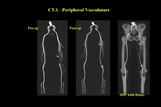
CT angiography is a technique that allows evaluation of the vasculature with a peripheral venous injection of contrast. This new application is possible because scanning can be completed during peak vascular enhancement. The fast scanning time allows the scan to be completed in a single breath hold, which greatly decreases motion artifacts. Additionally, the volumetric data set enables overlapping reconstructions, which facilitate construction of sophisticated three-dimensional renderings and multiplanar reconstructions. (Figure 7 & 8)
|

CTA - Peripheral Vasculature (Figure 7) |
|
The image shown here is an exceptional example of the scanning capabilities of the helical scanner and how it can be used to capture contrast in the peripheral vascular structures. Moving from left to right: The first image shows an occlusion of the deep femoral artery, proximal to the popliteal fossa. Collateral vessels are shown proximally to the occlusion which helped revascularize the left lower leg. Surgery was performed and a post-operative CT shows a patent femoral artery bilaterally. The image on the far right shows the power of image reconstruction by displaying both the vasculature and bone together. Relationship is very important because here you see the extent of surgery and internal landmarks. Follow-up exams will show patency of the vessels or recurring disease.
|

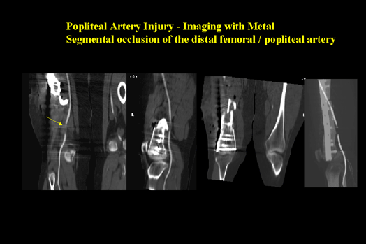
Popliteal Artery Injury (Figure 8) |
|
This helical CT exam shows an occlusion of the distal femoral/popliteal artery caused by traumatic injury and rod placement. After the helical exam is completed, a variety of multiplanar reconstructions was performed. Moving from left to right: Coronal reformat showing the full length of the vessel. Sagittal reformat showing the relationship of the vessel to the femur and the vessel. Coronal reformat showing the bony injury and screws. Sagittal maximum intensity projection displaying the rod and screws, bone and vessel.
|