序
六年前的今天(8/20/2015),发现得了肺腺癌,四期。经一年多的化疗,和后继的支持疗法,及种种努力,苟且至今。
https://blog.wenxuecity.com/myblog/73054/118522.html
正文
今年以来,身体、精神状态一直不太好,入夏后更糟。咳嗽得厉害,左胸疼痛加剧,己影响到日常活动和睡眠。
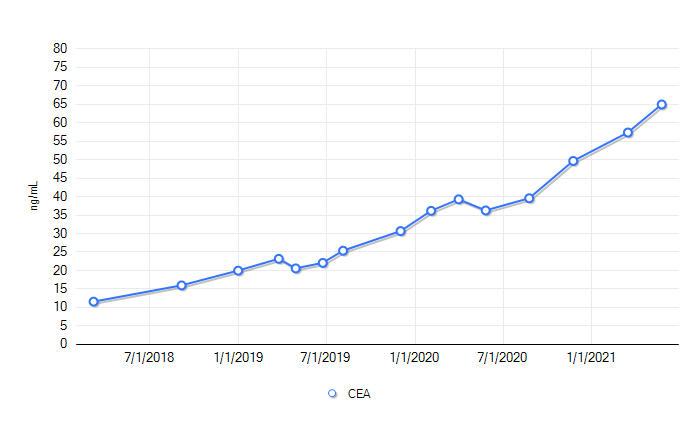
四月初做了CT,没看到啥变化。可CEA测值一直升高。
总觉得不对劲,跟医生要求做PET。
扫描结果证实几个热点。
真的,复发了,左肺内局部转移。六年前,在左下肺。这回在左上肺,除了淋巴转移,胸壁上也有,比较麻烦。不过仍然局限在左胸,身体其它部位没有热点。
8/3/2021 与A医生面谈,仔细看了扫描图片,並与四月初的CT片子和去年三月的PET扫描做了比较。单从影像角度,尺寸没有变化。PET扫描的热点(SUV),证实了恶性病灶的存在。医生也说,病灶小的时候,CT不是很灵。
又讨论了下一步诊治。确定了先做气管镜下细针穿刺活检病理和基因检测。再做脑MRI,根据检查结果,决定治疗方案。鉴于有胸壁多处转移,不考虑手术和放疗。
回家后,约了胸外科医生做镜检。这属于手术类操作程序,按现行规定,要求术前两天内新冠核酸检测阴性。
8/13/2021 周五 一大早去排车队,被捅了鼻子,双侧。
第二天早上收到报告:COVID-19 Test Result is Negative
8/17/2021 周二 早八点的镜检,要七点签到,一连串的问题要回答,一大堆文件要签字,一大群人来去接各种管线。迷过去之前,墙上挂表为八点十分。醒来时已在恢复室,九点半了。
换衣走人,还有些头晕,走不稳。护理推来轮椅,送我到楼下门厅。俺家总管已停车在门口等待。
到家后,躺在沙发休息,时不时咳口痰,带着血丝。也不敢睡,怕出血不止,如不及时……
中午,开始发烧,最高时38.8C。没服药,昏昏沉沉眯了一会儿。
下午六点,烧退了一些,在37.5。就咸鸡蛋喝了一碗白粥。
咳痰中仍有少许血丝。
晩上睡了一觉,早上起来已经不发烧了。咳痰中带着几小块凝血,没有新鲜血丝。
以为没事了,不料一阵剧咳后,痰里又有新鲜血丝了。赶紧吞了一片止咳药。
19号下午,胸外医生打来电话,说病理报道出来了,跟六年前一样,肺腺癌。他帮不了更多了,让我直接与A医生连系后继诊治。
8/28/2021 周六,早上做了脑MRI。
二天后收到报告:没发现转移灶
这是个好消息,肺癌最怕出现骨、脑转移。两者还没有,没到最坏的程度。
还在等外送的基因检测结果,如果配不上新药,再下一周,可能开始化疗,或其它疗法。
待下篇讲治疗
后注:
经历过了,流程熟了,略有紧张,不会焦虑。坦然面对,积极准备。
已经定购了各种辅助治疗的食材、中药、维生素、supplements ,这几天陆续收到快递。
购买了各种高蛋白低碳水食品,贮存在冰箱里。
本月初就开始服用能强化细胞免疫的东东了,但愿有帮助。
附注:
7/28/2021 扫描报告
THORAX:
Interval development of 1.2 cm FDG avid lymph node in the left
periesophageal region with SUV max of 4.6. Interval development of FDG
avid left hilar lymph node measuring 1.2 cm in diameter with SUV max of
3.3. Redemonstration of mildly prominent right upper paratracheal lymph
node measuring up to 1.1 cm that is similar to prior exam.
Interval increased FDG uptake along the medial pleural surface in the
left upper lung max SUV 3.3 previously 2.4. There is also diffuse
increased uptake in the remaining pleural surface slightly more focal
along posterior inferiorly (between left 10th and 11th ribs) with there
is corresponding soft tissue nodularity max SUV 3.6.
Impression
1. Compared to the previous PET-CT dated 03/24/2020 there is interval
development of FDG avid lymph nodes involving the left paraesophageal
region and left hilar regions. Findings are concerning for metastatic
lymphadenopathy.
2. There is interval increased FDG uptake along medial pleural surface
of the left upper lung and few other scattered posterior pleural
nodules including interval soft tissue nodule posterolaterally at level
of left 10th and 11th ribs. Findings are concerning for recurrent
disease.
肺左上肺内淋巴结细针活检病理报告结果:
| Comments: |
|
- Metastatic non-small cell carcinoma, favor adenocarcinoma.
Comment: The neoplastic cells are strongly and diffusely positive for TTF-1. This result supports the above rendered diagnosis. The control stain functioned appropriately.
|
脑MRI 报告
Impression
*No significant interval change since the previous examination. Stable
mild supratentorial white matter chronic microvascular ischemic changes.
*No acute intracranial findings or enhancing intracranial metastases.
下一篇:开打
|